Sunday 27 February 2011

Sketches I drew on a recent trip to a hospital

Some of these won't make much sense without commentary, but there are some that show clearly positions of electrodes on the body, how cables are sometimes stored and how hands may operate and untangle them.


Thursday 17 February 2011

Prep for expert interview

I am currently preparing my final questions for my expert interview tomorrow.  Although I am considering my questions, I believe it is important to keep things loose, questions will be answered and transformed, I may suddenly think of a question tomorrow that is relevant and have to shoe horn it into the conversation.

The thing is to keep the momentum of the conversation light and engaging.

In other news I found an interesting cable solutions company to speak to.

http://www.kalestead.co.uk/

I also need to speak to buildings and estates at Ninewells as soon as possible.

Friday 4 February 2011

Materials to use in making cultural probe

I am going to build a cultural probe, which is essentially a mockup of an ecg machine.  Staff and patients will be able to interact with it how they please, they can manipulate it by drawing on it, writing on it, turning it upside down, the results should be quite interesting.  I also plan to involve people who have never had an experience with an ecg machine before.  The pictures show a rough idea of what's to be built and some of the materials involved.  There are clips for electrodes on ecg machines so I am using clothes pegs and industrial type clamps to simulate these, people can then clip notes or other things to them, expressing their thoughts and opinions.

The pictures show rough sketches and materials to be used.


Friday 28 January 2011

co-ordinating with cardiographer

Before starting this semester I had the opportunity to observe the process of attaching and un-attaching a five lead electro-cardiogram (ecg) machine within the cathlab. During this day the cardiographer would describe to me what he would look out for on the monitors in his observation area.

I have just phoned the hospital and in the following week or two I should be able to shadow the cardiographer throughout his day in the cardio respiratory department. Although I have been to this department before it was essentially a tour of the machines and I had the opportunity to take photographs of the cables and wires as they lay.

This shadowing exercise will give me an insight into the actual processes and stresses the cardiographer encounters during the day especially in relation to tangled cables and wires.

Sunday 23 January 2011


For the master of design programme I have investigated the issues caused by poor facilities for cable management of an electro cardiogram (ECG) machine within the hospital environment.  Specifically the project research has drawn me to the cardio respiratory department of Raigmore hospital Inverness, shadowing staff and patients recording observations and their valuable insights.

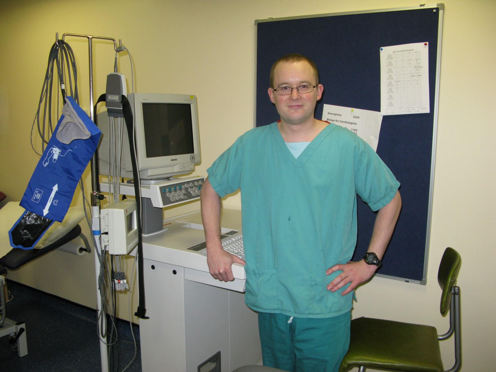
Ecg machines record the electrical activity of the heart and can involve as many as twelve wires attached via electrodes from the legs, arms and chest of the patient to a machine.  Due to time constraints and lack of proper storage equipment, these wires are often draped entangled over the top of equipment.  Untangling cables and wires is a frustrating task for the staff and is an unattractive sight for patients, affecting their confidence and security. 

A suitable solution to this problem would benefit both user groups, improving time management for staff and becoming aesthetically pleasing for patients.

I would like to continue this project after the masters programme.  It is incremental design and can continue to be developed and refined as technology progresses.

David has an undergraduate degree in three-dimensional design from Grays School of Art at the Robert Gordon University in Aberdeen.  

Thursday 11 November 2010

History of the electrocardiogram from 1600-2005

While the ecg machine has not technically been around since 1600 the theory behind it has.  This pdf notes key points in the progression of the ecg machine from theory to practice.  Apart from being a fantastic timeline going through the history of its development there are some incredible moments such as a professor using his own pet dog 'Jimmy' with paws in jars of saline to demonstrate new advancements, and the first oesophageal electrocardiogram was achieved with the aid of a professional sword swallower in 1906.  Fantastic advancements in medicine by fantastic means.

http://www.ecglibrary.com/ecghist.html